


Osteochondrosis is a widespread disease of the intervertebral discs, occurring in people of various age groups.Today it is diagnosed in more than 40% of young people under 35 years of age and, among the older category, it affects 9 out of 10 people.It has several stages of development and the earlier the pathology is detected, the easier and more effective the treatment will be and the lower the risk of developing dangerous complications.
What is osteochondrosis
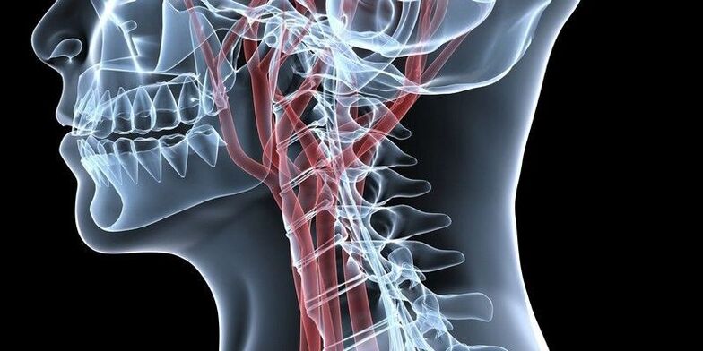
The intervertebral discs are located between the vertebral bodies and are a kind of shock absorbers that absorb stress when walking or performing other physical activities.They have different sizes depending on the position: in the cervical region the discs are the smallest and in the lumbar spine they are the largest.
They are all built the same way.In the center is the nucleus pulposus, which is the main component of the disc and has high elasticity.It is surrounded by a fibrous membrane and endplates.
Osteochondrosis is a degenerative-dystrophic disease in which a change in the shape and size of intervertebral discs occurs as a result of their abrasion and preconditions are created for the formation of hernias, deformation of the vertebral bodies, spondylosis and other disorders.
The thinning of the discs is a consequence of malnutrition of the cartilaginous tissue, which leads to a gradual decrease in its elasticity and an increase in fragility.
As a result, the discs change position, their height decreases and microcracks form in their fibrous membrane.This creates serious prerequisites for the formation of intervertebral hernias, compression of spinal roots or blood vessels.
The slightest changes in the condition of the disks lead to the interruption of their functions.This is accompanied by pain of varying degrees of intensity.At the same time, prerequisites for the development of diseases of internal organs are created, since the quality of their activity directly depends on the state of the spine and the conductivity of bioelectric impulses along the nerves.
It can affect one or more intervertebral discs anywhere in the spine.Therefore, osteochondrosis is diagnosed:
- cervical spine;
- thoracic spine;
- lumbar spine.
In especially severe cases, the pathological process involves most of the intervertebral discs of the spine, which is accompanied by discomfort in the entire back and almost complete loss of performance.But most often osteochondrosis affects the lumbar spine, as it bears the greatest load, as well as the intervertebral discs of the cervical spine due to their high mobility.
Development
During osteochondrosis, 4 stages can be roughly distinguished:
- Decrease in the hydration level of the disc, which leads to its dehydration and the formation of microcracks.Often, at this stage, there are still no manifestations of the incipient disease.
- Decrease in disc height, which leads to the appearance of the first symptoms of the pathology.At this stage, there is a decrease in the distance between individual segments, which leads to a decrease in the tone of the spinal ligaments and creates the possibility of displacement of the vertebrae from their natural positions, that is, the development of spondylolisthesis.In such situations, a wave-like course of the disease is most often observed.You can distinguish between periods of exacerbation, accompanied by acute pain, and periods of remission, in which there is no discomfort at all or there is intense pain.
- Deformation of the affected intervertebral disc with the formation of protrusion or prolapse.Sometimes there is involvement of the joints in the pathological process, which is manifested by the development of osteoarthritis or vertebral subluxations.At this stage, the immune system reacts to the processes occurring in the spine, developing aseptic inflammation with swelling of the tissues surrounding the affected spinal motor segment.In this case, pain occurs regularly, reflex muscle spasms can be observed, as well as mobility limitations.In rare cases, signs of neurological deficit (radicular syndrome) already develop as a result of compression of nerve fibers by protrusions resulting from the intervertebral discs.
- Development of complications.Since the human body has extensive compensatory capabilities, when the intervertebral disc loses its ability to perform its functions, the osteosynthesis process is activated to fix the vertebrae in a stable position.This leads to the formation of bony protrusions - osteophytes - at the edges of the vertebral bodies of the affected spinal motion segment.As a result, due to them, two or more vertebrae fuse tightly, forming a single conglomerate, that is, spondylosis develops.This always leads to compression of the nerves and the development of acute neurological symptoms in the form of paresis, paralysis and serious disturbances in the functioning of the corresponding internal organs.
Reasons
Today there are many theories for the development of osteochondrosis, including vascular, mechanical, hereditary, hormonal, infectious-allergic and others.But none of them are able to fully explain the mechanism of development of changes in the tissues of intervertebral discs.Therefore, most likely, they all complement each other.
Thus, a large number of very diverse factors can lead to the development of osteochondrosis.Among them, the most significant are:
- genetic predisposition;
- regular performance of heavy physical work;
- maintain a sedentary lifestyle;
- the presence of scoliosis or other spinal deformities;
- suffering from back injuries;
- unbalanced diet, vitamin deficiency;
- infectious diseases;
- metabolic disorders;
- congenital anomalies of the spine (Kimerli anomaly, Chiari anomaly, craniovertebral anomalies, sacralization, lumbarization);
- overweight;
- regular severe stress.
It is believed that the most significant influence on the condition of intervertebral discs is the constant overload of a certain mobile segment of the spine.This can be not only performing heavy and monotonous physical work, but also constant stooping or the habit of sitting for a long time.In these situations, additional stress is placed on the discs, muscles and ligaments, and other factors only worsen the situation.
In general, osteochondrosis can be considered an almost natural age-related disease, which is an inevitable price to pay for walking upright.
Symptoms
The first sign of the development of the disease is the appearance of a sharp sound in the part of the spine where the beginning of degenerative dystrophic changes is observed.It is a consequence of the occurrence of disturbances in the nutrition of the disc and the progression of its dehydration.At this stage, patients almost never seek medical help.Therefore, the daily routine, eating habits and other factors remain the same, which contributes to the worsening of the condition and the transition of the disease to the second stage.
As a result, typical symptoms of osteochondrosis begin to appear:
- severe muscle tension in the affected area on only one or both sides of the spine;
- sharp, aching pains that intensify with each movement and gain intensity over time, becoming unbearable;
- numbness in the arms or legs;
- weakness;
- stiffness of movements, unauthorized limitation of mobility;
- decreased muscle tone up to complete atrophy;
- poor posture;
- blood pressure spikes.
As the pathology progresses, signs of osteochondrosis worsen.80% of patients experience dull pain in the area of the affected spinal motion segment, which is also characteristic of myositis.
Osteochondrosis of the cervical spine
When the cervical spine is affected, the pain tends to radiate to the shoulder girdle, arms and other manifestations of neurological disorders occur.Patients keep their head in the least painful position and, if necessary, rotate their entire body.
In addition, pathologies of organs innervated by the spinal cord region of the cervical spine may develop:
- tinnitus and diseases of the ENT organs;
- dizziness;
- blurred vision;
- migraines;
- increase levels of irritability and anxiety;
- sleep problems;
- increased risk of allergic reactions;
- decreased levels of thyroid hormones.
In osteochondrosis of the cervical spine, vertebral artery syndrome and vegetative-vascular dystonia are often additionally diagnosed.
Infection of the cervical spine with osteochondrosis, especially when complicated by protrusions and intervertebral hernias, can cause compression of blood vessels.This is fraught with impaired blood supply to the brain, which can be accompanied by attacks of dizziness, loss of consciousness and even a stroke.
Osteochondrosis of the thoracic spine
In osteochondrosis of the thoracic region, which is rarely diagnosed, the pain occurs in the spine, at the level of the shoulder blades, and can intensify even during deep breathing.They are often felt behind the breastbone, which can be confused with heart disease.
When the spinal roots are pinched, the risk of diseases of the internal organs increases, in particular:
- bronchi and lungs (asthma, bronchitis, pneumonia, pleurisy);
- gallbladder and its duct, liver (cholecystitis, jaundice, fat absorption disorders);
- pancreas and duodenum (digestive disorders, pain in the left hypochondrium);
- adrenal glands, which affect the overall strength of the immune system and can provoke the development of allergies;
- kidneys (urinary disorders, chronic pyelonephritis, glomerulonephritis, etc.);
- pelvic organs (digestive disorders, gynecological, urological diseases, infertility).
Fact: damage to the intervertebral disc of the 7th thoracic vertebra by osteochondrosis can lead to the development of diabetes mellitus.
Osteochondrosis of the lumbosacral spine
With osteochondrosis of the lumbar region, low back pain, called lumbago, is observed.This is accompanied by burning, unbearable pain that occurs suddenly.Often, patients even have difficulty sitting, standing and walking, which may indicate the development of radicular syndrome.In these situations, it is easy to see how they sit down and stand up slowly, trying their best to avoid tilting their torso.
If complications occur, the main danger is compression of the cauda equina nerves, as this can impair control of the intestinal and bladder emptying processes, as well as paralysis of the legs.In these cases, the following may also occur:
- appendicitis;
- diarrhea, constipation;
- pain in the lower abdomen;
- bladder dysfunction;
- impotence;
- pain in the knees, feet, hips or groin area;
- swelling of the legs.
Complications
Osteochondrosis is a possible cause of a large number of different diseases.Most of the time, if left untreated, it leads to the formation of protrusion and intervertebral hernia.This, in turn, can cause:
- discogenic myelopathy, which ends in paresis, muscle atrophy, changes in tendon reflexes, loss of control over urination and defecation and even paralysis of the limbs;
- radiculopathy;
- scoliotic or other spinal deformity;
- spinal cord infarction due to compression of the artery that feeds it;
- stroke due to compression of the occipital artery.
Diagnosis
The appearance of back and neck pain should be a reason to consult a neurologist or vertebrologist.The earlier osteochondrosis is diagnosed, the easier and more effective the treatment will be.
To diagnose the disease, the doctor interviews and examines the patient.Based on their results, it is now possible to assume the presence of degenerative changes in the intervertebral discs.But for a final diagnosis, instrumental diagnostic methods are prescribed, including:
- magnetic resonance imaging;
- TC;
- X-ray in two projections.
MRI provides the most complete information about the condition of the intervertebral discs.The procedure is carried out mainly using closed devices with a power of 1.5 T. With its help, it is possible to differentiate osteochondrosis from tuberculous spondylitis, osteomyelitis, infectious diseases, etc.
Computed tomography and X-rays provide information about the bony structures of the spine.Thanks to them, it is possible to detect displacements of the vertebral bodies, the presence of osteophytes and other disorders.
Additionally, the following may be prescribed:
- Ultrasonography with Dopplerography of cervical vessels;
- electromyography;
- laboratory research.
Treatment of osteochondrosis
Therapy is developed individually for each patient.In this case, the severity of degenerative-dystrophic processes, the presence of complications, the nature of the patient's work activity and a number of other factors must be taken into account.
All patients must receive a prescribed set of measures, since it is impossible to eliminate pathological changes in intervertebral discs with the help of medications alone.Components of conservative therapy for osteochondrosis may include:
- drug therapy;
- osteopathy;
- manual therapy;
- physiotherapy (phonophoresis, ozone therapy, carboxytherapy, pressotherapy, RF currents);
- individual sessions with a rehabilitator.
All patients diagnosed with intervertebral disc dystrophy are advised to reconsider their lifestyle.It is essential to set aside time for moderate physical activity, especially for representatives of sedentary professions, or, on the contrary, think about the possibility of changing professions for people who are forced to lift heavy objects every day.
But in the acute period, complete rest is recommended.It is guaranteed not only by maintaining bed rest, but also by using orthopedic bandages: in case of injury to the cervical spine, a Shants collar is used;in case of osteochondrosis of the lumbar region, it is recommended to wear a corset.

In the early stages of the development of the disease, it is often enough to make lifestyle adjustments, practice exercise therapy and consult a chiropractor.In more advanced cases, drug therapy and physiotherapy are necessarily prescribed.
Never resort to self-medication with dubious means, self-prescription of anti-inflammatories, as well as dubious “healers” who position themselves on the Internet: you can lose not only time, money, but also the remains of your health.
Drug therapy
For osteochondrosis, a complex of medications is prescribed to reduce pain, eliminate inflammation and reflex muscle spasms, improve nerve conduction and activate the tissue regeneration processes of intervertebral discs.
Therefore, patients are prescribed:
- NSAIDs – help reduce pain and have an anti-inflammatory effect;
- corticosteroids – have powerful anti-inflammatory properties;
- muscle relaxants – eliminate muscle spasms, which helps reduce back pain;
- B vitamins – provide better functioning of the nervous system in general and the conduction of nerve impulses along individual nerves in particular;
- vitamin D is a medicine responsible for the state of bone tissue, as well as higher brain functions, such as memory, attention, speech;
- chondroprotectors – nourish the intervertebral discs with the compounds necessary for the construction of new fibers of the nucleus pulposus;
- psychotropic medications – increase the effectiveness of non-steroidal anti-inflammatory drugs and muscle relaxants;
- vascular medicines - improve blood circulation in the tissues surrounding the spine, which ensures a more active supply of nutrients and oxygen to the intervertebral discs;
- Anticonvulsants – used in rare cases to relieve very severe spasms.
For very severe pain, which most often indicates complications, patients can undergo back blocks, which take effect immediately.Sometimes corticosteroid hormones are added to the solution to accomplish the blockage.Furthermore, this leads to a pronounced anti-inflammatory effect.
Blocks are carried out under conditions of absolute sterility, which can only be achieved in specialized medical institutions.At the same time, they require special knowledge and skills, so only a highly qualified healthcare professional can carry out the task competently.Otherwise, there is a high risk of infection or damage to the nerve fiber, which will lead to the development of serious complications.
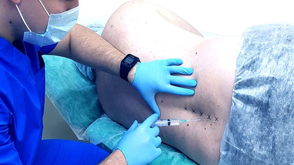
During the block, injections are usually given to both sides of the spine, in the area where the nerves that cause the pain occur.There are several techniques to perform them, among which the specific one is selected individually by the doctor.
But it is advisable to carry out blocking no more than 4 times a year.Since in osteochondrosis, acute and aching pain attacks occur much more frequently, it is first of all worth directing efforts to eliminate the causes of their occurrence, that is, the degenerative-dystrophic process in the joint itself.
Manual therapy
Manual therapy sessions are prescribed outside the period of exacerbation of osteochondrosis.They play one of the main roles in the treatment of the disease, since the competent use of manual techniques on the spine and surrounding tissues helps not only to stop the progression of the pathological process in the intervertebral discs, but also to create the most favorable conditions for its restoration.
Furthermore, certain techniques can also have a positive effect on the functioning of internal organs.For example, one of the methods is designed, by normalizing the position of each vertebra, to eliminate pressure on the spinal roots, blood vessels and spinal membranes and thereby restore the normal connection of the organ with the nervous system.This allows you to eliminate the hidden causes of the development of the above-mentioned diseases of the heart, bronchi, lungs, kidneys, gastrointestinal tract and reproductive system and leads to complete recovery.
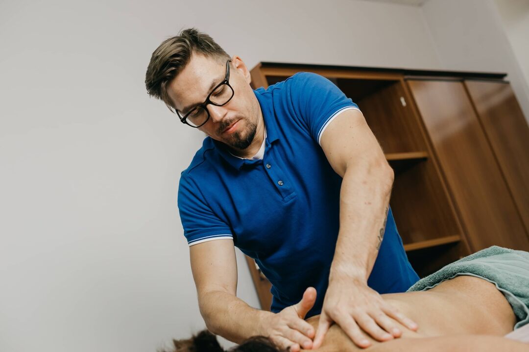
Thanks to the precise effect of the jewel on the spine, not only is blood circulation activated and metabolism accelerated, as happens with a classic therapeutic massage, but natural self-healing mechanisms are also launched.Eliminating curvatures of the spine, incorrect position of the vertebrae and other pathological changes in the spine through manual therapy can further strengthen the immune system, improve overall well-being and significantly improve the quality of life.
Positive changes can be noticed after the first session, and in the future their severity only increases.
Physiotherapy
Physiotherapeutic procedures increase the effectiveness of all other methods of treating osteochondrosis and help reduce pain.Most often used:
- electrophoresis - use of electric current to ensure the penetration of anesthetics, anti-inflammatory drugs and other agents directly into the site of inflammation, allowing a pronounced therapeutic effect to be quickly obtained;
- Ultrasound therapy - the effect of ultrasound provides an analgesic effect, increases the intensity of metabolic processes in the area of influence and creates the prerequisites for high-quality restoration of thinned intervertebral discs;
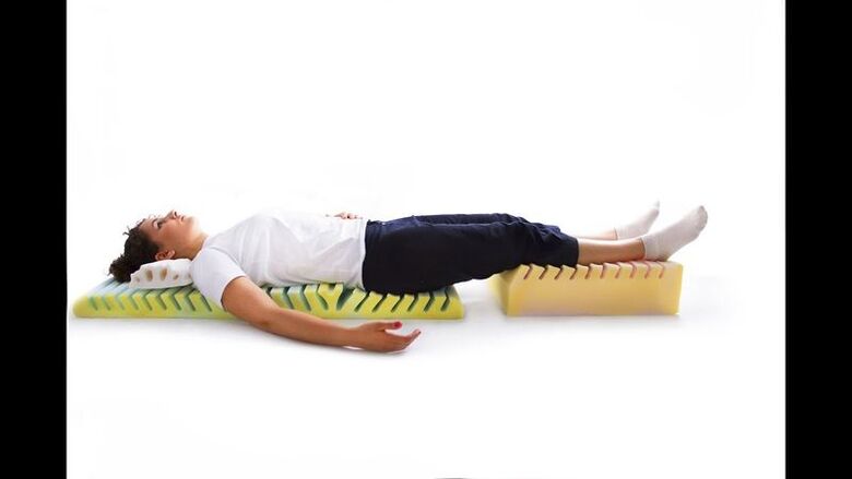
- detensor therapy – involves stretching and massaging the spine on a special ribbed mattress, which activates blood circulation and increases muscle tone;
- traction therapy or spinal traction – is carried out using special devices that create a traction load on the spine, as a result of which the distance between the vertebrae increases and the pressure on the worn out intervertebral disc decreases sharply, which allows for more active recovery.
All procedures are prescribed in courses of 10 to 15 sessions.When choosing them, they take into account not only the state of the intervertebral discs, but also the presence of concomitant diseases, since in some of them certain physical effects are contraindicated.
Exercise therapy
Therapeutic exercises for osteochondrosis play a significant role, since correctly selected exercises for injuries in different parts of the spine can help form a strong muscular corset and at the same time eliminate increased muscle tone.This will provide good support for the spine and stop the progression of the degenerative process in the intervertebral discs.In addition, exercise therapy helps to activate blood circulation and increase the intensity of metabolic processes.
Developing a physiotherapy program is the task of the rehabilitation doctor.Based on the severity of the spinal cord injury, the characteristics of the patient's physical development, his age and other factors, he will create an optimal set of exercises, the implementation of which will create an optimal load on the muscles and spine.
The first classes must be held under the supervision of a doctor.After the patient learns to perform each exercise correctly, he or she can continue practicing at home.It is important to avoid sudden movements.All exercises are performed daily slowly and smoothly, and the load is gradually increased.But the occurrence of pain during exercise is a good reason to refuse to perform the exercise that caused it.

Prevention
It is much easier to prevent the development of a disease than to deal with its consequences later.Since osteochondrosis can occur in everyone sooner or later, you should think about possible risks as early as possible and make every effort to avoid them.Therefore, each person must adhere to the following recommendations:
- avoid physical inactivity, exercise regularly, swim and, during sedentary work, take regular breaks to warm up;
- pay attention to your posture when walking or sitting;
- purchase a high-quality orthopedic mattress and pillow;
- observe the correct weightlifting technique: with your back straight and knees bent;
- swap casual shoes for more comfortable ones and leave dress shoes for special occasions;
- Eat well so that your body does not suffer from nutritional deficiencies and your weight remains within normal limits.
There is only one way to prevent the appearance and manifestation of osteochondrosis symptoms: start taking care of yourself and your health.Now osteochondrosis is not just a disease, but a complex of musculo-tonic and neurodystrophic changes, which are the body's response to physical inactivity, chronic static tension of the spinal muscles, the environmental situation and chronic neurosis, especially common among city residents, swimming in the pool and stretching programs, including yoga.
Thus, osteochondrosis is a very common disease, but with close attention to your health, you can effectively fight it at any age.But ignoring the problem will not lead to anything good and sooner or later will force the patient to lie down on the operating table.